
Objective evidence of ligament damage (not AOMSI)
- claytonchiropractic
- 2 hours ago
- 4 min read
For the first 3 podcasts I covered a lot of information on ligament damage. The second podcast in particular I spent a lot of time talking about alteration of motion segment integrity or AOMSI. That podcast shows how we can objectively show catastrophic ligament damage. As a refresher AOMSI is when we have changes in the angle between the motion segments in the spine of over 11 degrees. AOMSI is also when we have translation forward or backwards of the vertebra by 3.5mm or 20% of the vertebra.
Once we see AOMSI on the x-rays we know there is catastrophic ligament damage. My 1st and 3rd podcast discuss what this means to patients. For today’s podcast I want to cover ligament damage that is below the threshold for AOMSI or what I call sub catastrophic ligament damage. Today’s study comes from the Spine Journal in 2001 titled Characteristics of Sagittal Vertebral alignment in Flexion determined by Dynamic radiographs of the cervical spine by Ruey-Mo Lin et al.
This study we attempting to quantify the wide variation of normative values of change in angular motion and translation in the cervical spine with flexion and extension x-rays. Or as they said quote “ to investigate the normal behavior of cervical spine” end quote.
For this study they found a population of patients who’s spines qualify as normal. They included only patients who never had neck pain that limited the patients work or recreational activities or that caused them to seek medical help. They also excluded any patients that had x-rays showing kyphosis, scoliosis, spondylolisthesis and fusion.
They took this group of normal patients and took lateral x-rays in neutral, flexion and extension. They then measured angular displacement and translation. Of note 10 subjects there was no data for C6-7. This was due to this level not being visible on the x-rays. The lower cervical spine can be difficult to image in some patients due to size. On a lateral view the lower cervical spine is often at the level of the shoulders making imaging difficult.
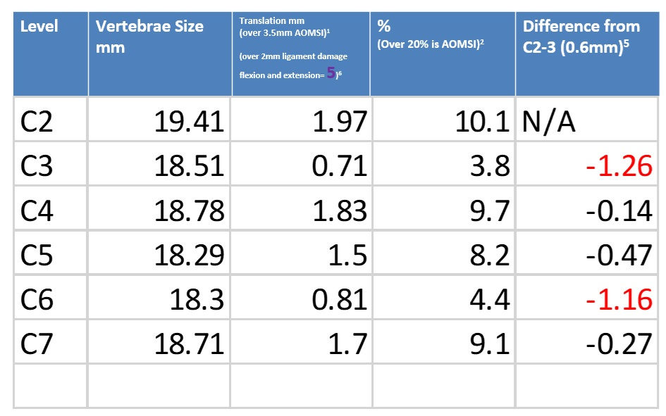
This study was able to find normal levels for healthy populations of angular displacement and translation. They used each patient’s x-rays as a baseline to measure off of. Given the stability of C2-3 they used this level as the baseline. This is different than the AOMSI study which used adjacent levels as the baseline. This study uses C2-3 as the baseline since quote “most of the trauma, degenerative changes, and instability occur in the lower cervical spine.” End quote.
Quote “When C2-3 was used as a baseline to calculate the intervertebral difference of angular displacement and translation in the flexion position, nearly all the intervertebral differences of angular displacement were less than 7 degrees, and those of translation were less than 0.6mm” end quote.
They reported that they used self-comparison measurements given the wide variations in total flexibility. This study is very simple, but very powerful. With the AOMSI study we can objectively show catastrophic ligament damage. With today’s study we can show objective evidence of ligament damage that places the patient outside of normal.
Patients that fit the criteria of today’s study have a level of ligament damage that can objectively be shown to no longer fit in the healthy population. As seen in the first 3 podcasts ligaments undergo micro tearing or complete tearing of ligaments. This study shows that patients did not have complete tearing, but enough tearing to alter the spine out of what we see in the normal population.
This type of ligament damage will to an extent qualify for all the changes covered in the first 3 podcasts. As a refresher these damage ligaments have horrible and incomplete healing. They will be replaced with inferior collagen, fat cells, blood vessels and scar tissue. This inferior healed ligament will alter the mechanics of the spine leading to degenerative bony changes. The reflexive control the ligaments have over muscles will to an extent be altered with this ligament damage.
To recap today’s study we are talking about taking flexion and extension x-rays of the spine to assess for ligament damage. If the x-rays do not show AOMSI with change in angular motion of 11 degree compared to adjacent segments or 3.5mm translation or 20% translation then they do not have AOMSI or catastrophic ligament damage. Todays study can objectively show ligament damage, but not the catastrophic level, but at a level where the patient is not longer comparable to the healthy population.
This can be done by using only flexion x-rays to see if any level of angular displacement is 7 degrees more than C2-3. Once we see angular displacement 7 degrees or more above C2-3 then we know we have damage to these ligaments. This is not seen on extension x-rays, but just the flexion.
For translation we use both flexion and extension x-rays. If we see translation 0.6mm more than C2-3 then we know we have ligament damage and this patient no longer fits in the normal population. These are simple measurements that can be done to objectively show if damage was done to the cervical spine.
Real world:
For today’s real world example, I would like to have guest on the podcast. Someone who has personal experience of this type of damage.


Comments